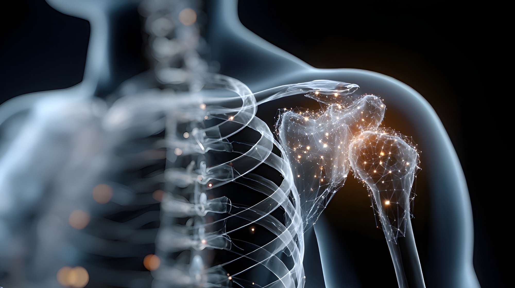


Common Shoulder Cartilage Injury Symptoms & Causes
Shoulder cartilage injury symptoms vary depending on damage severity and location but typically include deep aching pain localized within the joint, worsening with overhead activities or lifting, mechanical symptoms such as catching, locking, or grinding sensations during movement, stiffness particularly after periods of rest, swelling and effusion following activity, reduced range of motion especially with rotation and elevation, weakness affecting functional activities, night pain disturbing sleep particularly when lying on the affected shoulder, and progressive symptom deterioration over time. Small superficial lesions may cause minimal symptoms initially while large full-thickness defects create significant disability. Loose cartilage fragments or flaps can cause sudden sharp pain with certain movements and mechanical catching requiring arthroscopic removal.
Causes of shoulder cartilage damage include traumatic shoulder dislocations—the most common cause of acute cartilage injury, often creating chondral defects or osteochondral fractures on the humeral head or glenoid; repetitive overhead activities in throwing athletes, swimmers, and manual laborers causing cumulative microtrauma and degeneration; previous shoulder injuries including fractures healing with joint surface irregularity; chronic shoulder instability with recurrent subluxations damaging cartilage over time; rotator cuff tears causing abnormal joint mechanics and secondary cartilage wear; inflammatory arthritis including rheumatoid arthritis directly attacking cartilage; avascular necrosis of the humeral head causing cartilage collapse; post-surgical changes following previous shoulder operations; and age-related degeneration as cartilage naturally degrades with aging particularly after age 40.
Schedule Your ConsultationWho's at risk for Shoulder Cartilage Injuries?
Multiple factors increase susceptibility to shoulder cartilage damage. Identifying risk factors enables early intervention and prevention strategies:
Athletes and Overhead Activity
+Overhead athletes face dramatically elevated risk from repetitive microtrauma. Baseball pitchers, particularly those throwing high pitch counts without adequate rest, develop cartilage degeneration from repetitive rotational stress. Swimmers performing thousands of strokes weekly, volleyball players, tennis players, and javelin throwers experience similar cumulative damage. Manual laborers including painters, construction workers, and warehouse employees performing repetitive overhead work sustain occupational cartilage injuries. Weekend warriors suddenly increasing activity intensity without proper conditioning risk acute injuries.
Previous Shoulder Injuries and Instability
+Prior shoulder dislocations dramatically increase cartilage injury risk, with studies showing chondral damage in over 40% of first-time dislocations and higher rates with recurrent instability. Previous fractures involving joint surfaces, particularly those healing with step-off or incongruity, cause abnormal contact pressures accelerating cartilage degeneration. Chronic shoulder instability from labral tears or ligament laxity creates repetitive abnormal joint loading patterns damaging cartilage progressively. History of shoulder surgery, particularly if joint mechanics weren't fully restored, increases secondary cartilage wear risk.
Rotator Cuff Pathology and Biomechanical Factors
+Rotator cuff tears alter shoulder biomechanics causing superior humeral head migration and abnormal contact patterns that accelerate cartilage breakdown. Larger tears and those present longer create more severe cartilage damage. Scapular dyskinesis and poor shoulder blade control cause abnormal joint positioning during movement increasing cartilage stress. Previous rotator cuff repairs that don't fully restore anatomy may leave residual mechanical abnormalities promoting cartilage degeneration. Muscle imbalances and weakness compromise joint stability and protection.
Age and Degenerative Factors
+Age is a primary risk factor with cartilage naturally degenerating over time. Individuals over 40 show increasing cartilage changes with age-related breakdown accelerating after 50. Genetic predisposition influences cartilage quality and degeneration rate with family history of early arthritis indicating increased risk. Inflammatory conditions including rheumatoid arthritis, lupus, and other autoimmune diseases directly attack cartilage causing progressive destruction. Metabolic conditions like diabetes and gout can affect cartilage health. Obesity increases joint loading accelerating wear. Smoking impairs cartilage nutrition and healing. Avascular necrosis from corticosteroid use, alcohol abuse, or other causes leads to cartilage collapse.
Shoulder Cartilage Injury Prevention
Prevention focuses on reducing cartilage stress and maintaining joint health. For overhead athletes, implement proper training programs with adequate rest periods allowing cartilage recovery, pitch count limitations for baseball players, graduated return to sport after layoffs, sport-specific conditioning strengthening shoulder stabilizers, proper technique instruction reducing abnormal joint stress, and cross-training avoiding excessive repetitive movements. Address shoulder instability promptly—unstable shoulders should be stabilized surgically before progressive cartilage damage occurs. Treat rotator cuff tears appropriately as untreated tears cause secondary cartilage degeneration. Maintain shoulder strength and flexibility through regular exercise focusing on rotator cuff strengthening, scapular stabilizer exercises, and full range of motion preservation.
Control modifiable risk factors including maintaining healthy weight reducing joint loading, avoiding smoking which impairs cartilage nutrition, limiting alcohol consumption, managing inflammatory conditions with appropriate medical treatment, controlling diabetes and metabolic disorders, and using corticosteroids judiciously given avascular necrosis risk. For those with previous shoulder injuries, complete rehabilitation ensuring full strength and motion restoration before returning to activities. Use proper ergonomics for repetitive overhead work with periodic position changes and strengthening breaks. Address early symptoms promptly—catching, grinding, or persistent pain warrant evaluation before damage progresses. Once significant cartilage damage exists, prevention is no longer possible—focus shifts to preventing progression through activity modification, joint protection, and appropriate treatment. Early identification and intervention when cartilage damage is less severe yields better outcomes than waiting until advanced degeneration develops.
How are Shoulder Cartilage Injuries Diagnosed?
Diagnosis begins with detailed history documenting pain location and quality (deep aching versus sharp catching pain suggests different pathology), activity limitations particularly overhead activities and lifting, mechanical symptoms including catching, locking, or grinding, previous shoulder injuries or dislocations, prior shoulder surgeries, athletic or occupational demands involving overhead work, and symptoms of inflammatory conditions. Physical examination includes inspection for muscle atrophy suggesting chronic rotator cuff pathology, palpation assessing tenderness localized to joint line, range of motion assessment documenting limitations and painful arcs, strength testing evaluating rotator cuff function, stability testing for instability contributing to cartilage damage, and special tests including impingement signs and load-and-shift tests. Provocative maneuvers may reproduce mechanical symptoms.
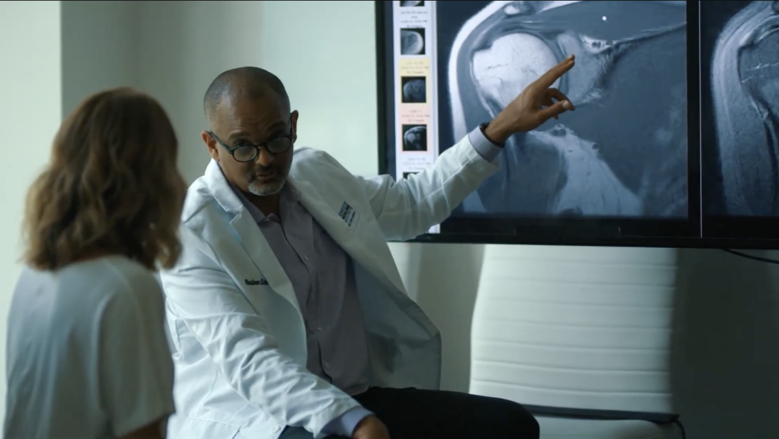
Standard X-rays evaluate joint space narrowing indicating cartilage loss, subchondral bone changes including sclerosis or cysts, osteophyte formation suggesting degenerative changes, previous injuries or surgical changes, and bone alignment and joint congruity. Advanced imaging provides detailed cartilage assessment. MRI with specialized cartilage sequences shows cartilage thickness and integrity, identifies full-thickness defects and areas of thinning, evaluates for bone marrow edema indicating stress, assesses associated pathology including labral tears and rotator cuff damage, and helps grade cartilage injury severity. MR arthrography with intra-articular contrast improves sensitivity for detecting cartilage flaps and partial-thickness defects. CT arthrography offers alternative detailed assessment particularly for bony pathology. Diagnostic arthroscopy remains gold standard for definitive cartilage assessment, allowing direct visualization of lesion size and location, grade determination using International Cartilage Repair Society (ICRS) grading system, probing cartilage to assess stability and depth, identification of unstable flaps requiring debridement, and simultaneous treatment during diagnostic procedure.
What treatment is best for Shoulder Cartilage Injuries?
Treatment depends on cartilage defect characteristics including size, depth, and location, patient factors such as age and activity level, symptom severity and functional limitations, and presence of associated shoulder pathology. Treatment goals include relieving pain, improving function, preventing progression, and delaying or avoiding joint replacement. Options range from conservative management to advanced cartilage restoration procedures. Treatment is individualized based on comprehensive evaluation and shared decision-making.
Conservative Management and Activity Modification
+Initial treatment for many cartilage injuries focuses on conservative management including activity modification avoiding painful overhead activities and heavy lifting, physical therapy emphasizing rotator cuff and scapular strengthening to optimize joint mechanics, range of motion exercises preventing stiffness, NSAIDs for pain and inflammation control, and intra-articular corticosteroid injections providing temporary symptom relief particularly for inflammatory components. Viscosupplementation with hyaluronic acid injections may provide lubrication and symptom relief though evidence for shoulder is less robust than for knee. Nutraceuticals including glucosamine and chondroitin may offer modest benefit. Conservative treatment is appropriate for small superficial lesions, older low-demand patients, and those with minimal symptoms.
Arthroscopic Debridement and Marrow Stimulation
+Arthroscopic surgery addresses symptomatic cartilage lesions. Chondroplasty involves debriding unstable cartilage flaps, smoothing irregular surfaces, and removing loose bodies causing mechanical symptoms, providing symptom relief though not regenerating cartilage. Microfracture, the most common marrow stimulation technique, creates small holes in exposed subchondral bone allowing bone marrow stem cells and growth factors to form fibrocartilage filling the defect. Best suited for small to medium full-thickness defects (<2-4 cm²) in younger patients. Results include pain reduction and improved function though fibrocartilage formed is mechanically inferior to native hyaline cartilage. Success depends on defect size, location, and patient compliance with rehabilitation.
Advanced Cartilage Restoration Procedures
+Larger cartilage defects or failed previous treatments may benefit from advanced restoration. Autologous chondrocyte implantation (ACI) involves harvesting patient's own cartilage cells arthroscopically, culturing cells in laboratory for 4-6 weeks to expand numbers, then reimplanting cells under a membrane covering the defect where they regenerate hyaline-like cartilage. Requires two surgeries but produces more durable hyaline-like repair than microfracture. Matrix-assisted chondrocyte implantation (MACI) uses scaffold seeded with cells simplifying technique. Osteochondral autograft transfer (OATS/mosaicplasty) harvests cylindrical plugs of healthy cartilage and bone from non-weight-bearing areas of the shoulder and transfers them to fill defects, providing immediate hyaline cartilage surface. Best for medium-sized focal defects. Osteochondral allograft transplantation uses donor tissue for larger defects where autograft insufficient.
Shoulder Replacement for Advanced Damage
+Extensive cartilage loss with end-stage arthritis unresponsive to conservative treatment and cartilage restoration may require shoulder replacement. Total shoulder arthroplasty replaces both humeral head and glenoid with prosthetic components, providing excellent pain relief and function restoration for glenohumeral arthritis with intact rotator cuff. Reverse total shoulder replacement indicated when severe arthritis accompanied by rotator cuff deficiency, using different biomechanics allowing deltoid to power arm movement. Hemiarthroplasty replaces only humeral head, considered for younger patients with humeral-sided disease and preserved glenoid. Shoulder replacement is definitive treatment for advanced arthritis but reserved for older patients or those who have exhausted other options given implant longevity limitations and activity restrictions.
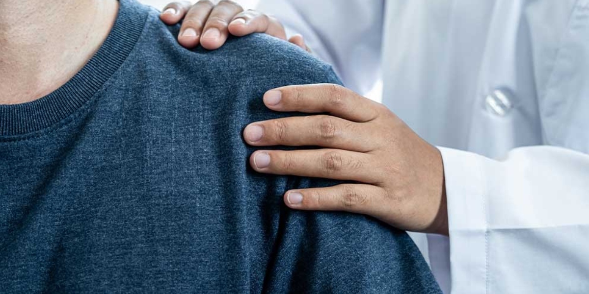
Shoulder Cartilage Injury Treatment in Cleveland, Ohio

Cleveland Shoulder Institute specializes in comprehensive evaluation and treatment of shoulder cartilage injuries utilizing the latest diagnostic imaging and treatment techniques. Our fellowship-trained shoulder specialists have extensive experience managing cartilage damage from early-stage lesions to advanced arthritis.
We offer complete cartilage injury assessment including high-resolution MRI with specialized cartilage sequences, CT arthrography for detailed joint imaging, and comprehensive physical examination evaluating all factors contributing to cartilage stress. Conservative treatment includes targeted physical therapy programs, regenerative medicine options, and activity modification guidance. For symptomatic cartilage lesions requiring surgery, we perform advanced arthroscopic procedures including precise chondroplasty, microfracture techniques optimized for shoulder anatomy, and removal of loose bodies and unstable flaps. We have expertise in advanced cartilage restoration including autologous chondrocyte implantation, osteochondral grafting, and complex reconstruction procedures. For patients with advanced arthritis, we offer shoulder replacement surgery with extensive experience in both anatomic and reverse total shoulder arthroplasty. Our treatment philosophy emphasizes joint preservation when possible, using biological treatments and cartilage restoration to delay or avoid replacement. Each patient receives individualized treatment based on cartilage damage characteristics, functional goals, and long-term joint health. Located in Cleveland with comprehensive follow-up ensuring optimal recovery and preventing progression.
Schedule Your ConsultationMeet our Shoulder Cartilage Injury Team
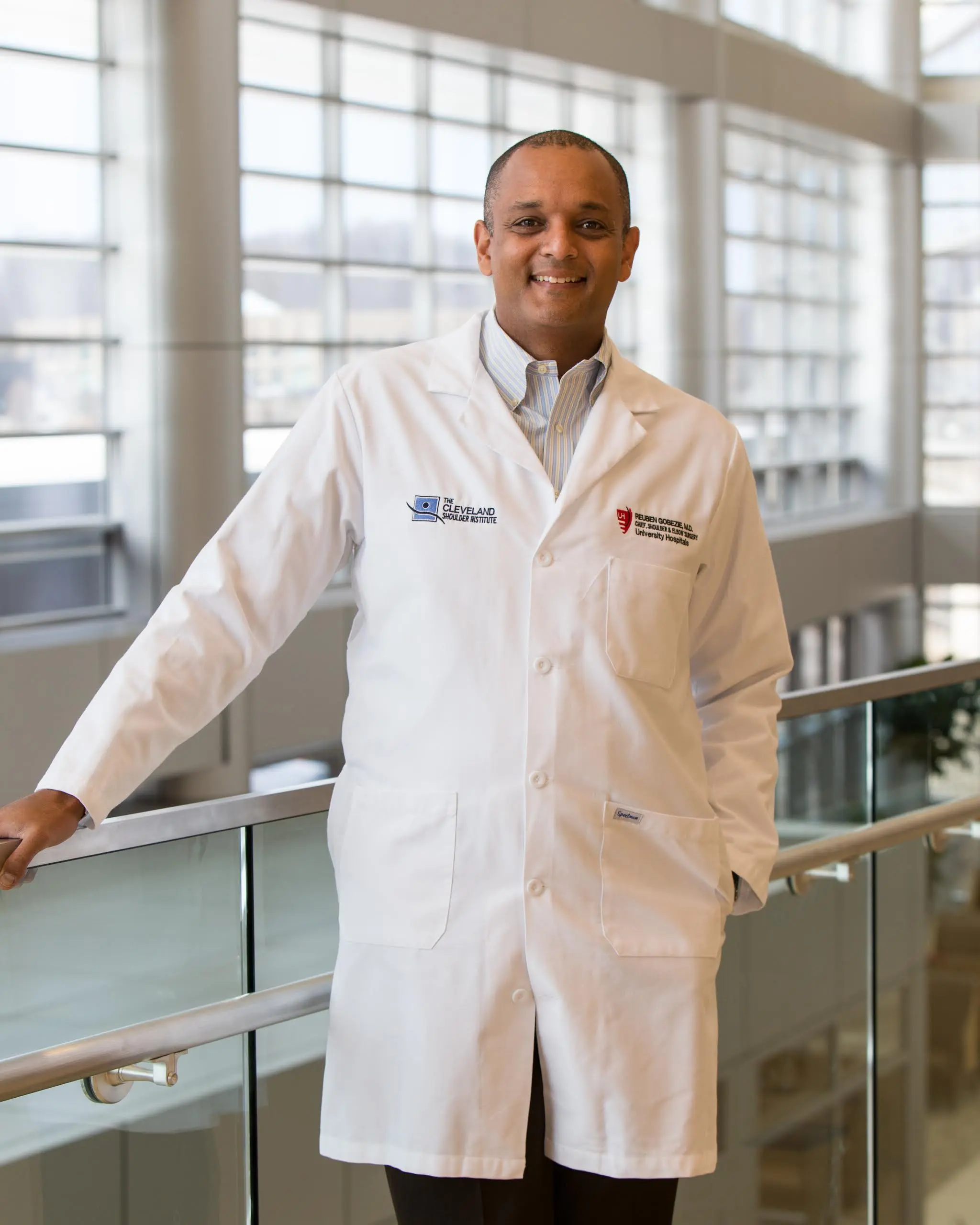
Dr. Gobezie is a fellowship-trained shoulder and elbow surgeon with specialized expertise in shoulder cartilage pathology and restoration. He has advanced training in arthroscopic cartilage procedures, biological treatments, and complex cartilage restoration techniques. Dr. Gobezie maintains expertise through active participation in cartilage research, teaching commitments training other surgeons in advanced techniques, and attendance at specialized shoulder conferences focusing on cartilage treatment innovations.
Supporting Dr. Gobezie are board-certified anesthesiologists, experienced surgical teams trained in arthroscopic and open shoulder procedures, specialized physical therapists understanding cartilage healing and joint preservation exercises, and dedicated medical staff. This collaborative approach ensures accurate diagnosis combining clinical evaluation with advanced imaging, appropriate treatment selection matching pathology severity to intervention, expert surgical technique when procedures are needed, structured rehabilitation optimizing cartilage healing and joint mechanics, and long-term monitoring preventing progression. Our team understands cartilage injuries significantly impact quality of life and athletic performance, providing compassionate care while offering cutting-edge treatments to restore function and preserve joint health.
What Our Patients Say About Cartilage Injury Treatment
Real experiences from patients who successfully treated shoulder cartilage injuries:
"As a competitive swimmer, shoulder pain from cartilage damage was ending my career. Dr. Gobezie performed microfracture surgery and guided my rehabilitation expertly. Eight months later I'm back competing at a high level. His specialized knowledge of cartilage injuries made all the difference."
— Rachel Thompson
"I had cartilage damage from an old shoulder dislocation that bothered me for years. Dr. Gobezie explained my options thoroughly including debridement versus cartilage restoration. We chose arthroscopic debridement and the results exceeded expectations. The mechanical symptoms resolved and I have much better function now."
— Michael Chen
"Dr. Gobezie diagnosed significant cartilage loss from arthritis. He was honest that I needed shoulder replacement given the damage extent. The surgery went perfectly and I have no pain now with great motion. Appreciate his expertise and straightforward communication about my condition and treatment options."
— Patricia Collins
Shoulder Cartilage Injury Frequently Asked Questions
Can shoulder cartilage heal on its own?
+Shoulder cartilage has very limited healing capacity because it lacks blood supply. Small superficial cartilage injuries may remain stable without progression but don't truly heal. Full-thickness cartilage defects exposing bone cannot heal naturally. This is why treatment focuses on stimulating repair through procedures like microfracture bringing blood supply to the area, or restoring cartilage through grafting or cell-based techniques. Early intervention when damage is limited offers better results than waiting.
What is the difference between microfracture and cartilage transplantation?
+Microfracture creates small holes in bone allowing blood and stem cells to fill defects with fibrocartilage—a repair tissue that is mechanically inferior to native hyaline cartilage but provides symptom relief and function improvement. Best for smaller defects in younger patients. Cartilage transplantation procedures like autologous chondrocyte implantation or osteochondral grafting restore actual hyaline or hyaline-like cartilage providing more durable long-term results. These are more complex procedures reserved for larger defects or failed microfracture. Your surgeon recommends treatment based on defect characteristics and patient factors.
Will I develop arthritis after cartilage injury?
+Cartilage injury increases arthritis risk though not all injuries progress to severe arthritis. Small stable lesions may never cause problems. Larger full-thickness defects, particularly those left untreated, often progress to arthritis over years. Risk factors for progression include defect size, location in high-load areas, associated instability or rotator cuff pathology, and patient age and activity level. Appropriate treatment including activity modification, cartilage restoration procedures, and addressing contributing factors can slow or prevent arthritis development. Early intervention offers best chance of preserving joint health long-term.
How long is recovery after cartilage restoration surgery?
+Recovery varies by procedure type. Arthroscopic debridement typically allows return to light activities in 6-8 weeks. Microfracture requires longer protection—immobilization for 2-3 weeks, physical therapy for 3-6 months, return to sports at 6-9 months allowing fibrocartilage formation. Autologous chondrocyte implantation requires extended recovery with limited weight-bearing for 6-8 weeks, progressive strengthening for 6-12 months, and sports return at 12-18 months while transplanted cells mature. Compliance with rehabilitation protecting repair while gradually loading tissue is critical for success. Your surgeon provides specific timeline based on procedure performed.
Can I continue playing sports with cartilage damage?
+This depends on injury severity and symptoms. Small stable lesions causing minimal symptoms may allow continued participation with activity modification and monitoring. Moderate lesions causing pain or mechanical symptoms often require treatment before safe sports return. Large full-thickness defects or progressive symptoms warrant treatment to prevent further damage. Continuing aggressive overhead sports with significant cartilage injury risks progression to more severe damage potentially requiring joint replacement. Your surgeon evaluates injury characteristics, symptoms, sport demands, and treatment options to guide activity recommendations and optimize long-term shoulder health.